Cruciate Disease: What it is and how it's treated

- posted: Feb. 08, 2024
WHAT IS CRUCIATE DISEASE?

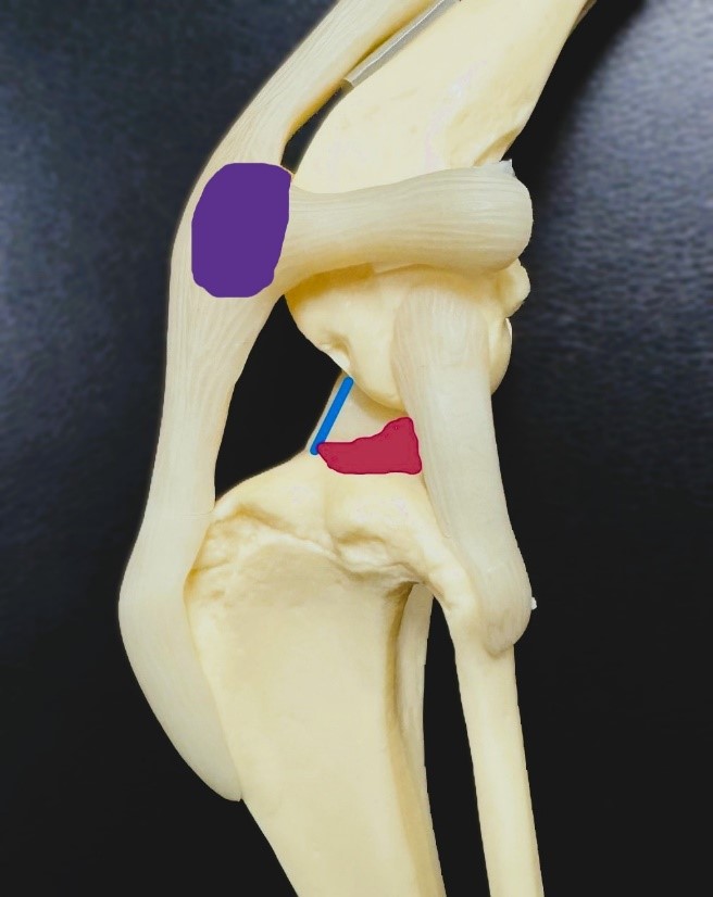
Canine knee joint – purple oval is the knee cap, blue line is orientation of the CrCL, pink wedge is the meniscus
In animals, the ACL is referred to as the Cranial Cruciate Ligament or CrCL. The CrCL is one of the ligaments important to stabilizing the knee and preventing the tibia (shin bone) from moving too far forward when walking/running. This ligament can either become suddenly torn due to injury (an acute tear, more common in small breed dogs and cats) or gradually fray and break down over time (chronic cruciate disease, more common in large breed dogs). Sometimes, the medial meniscus (one of the cushions in the knee) can also be torn, leading to more severe pain and lameness. For pets with acute CrCL tears, pain and lameness is usually sudden and severe. For dogs with chronic cruciate disease, they commonly only show “soreness” after strenuous exercise early in the disease. Over time, they either gradually become more and more sore and “slower” as joint inflammation and arthritis develops, or can become suddenly lame and painful if a large portion of the ligament or the meniscus suddenly tears. If left untreated, CrCL disease and injuries can cause significant arthritis, pain, and lameness. Once CrCL disease is affecting one leg, the pet will start weight shifting and putting more pressure on the other rear limb, increasing the likelihood of this leg also experiencing CrCL disease. It should be noted that in some animals with acute cruciate tears, their lameness may improve with time. This does not mean their ligament has healed or they don’t require surgery. Their comfort level initially improves as the joint swelling improves, but without intervention, arthritis will occur and cause worsening pain and lameness later on.
HOW IS CRUCIATE DISEASE DIAGNOSED?
Cruciate disease is often suspected and diagnosed with a combination of specific signs found on physical exam by a veterinarian and radiographs (x-rays) to evaluate arthritic changes and soft tissue inflammation in the knee joint. Definitive diagnosis is obtained by direct visualization of the CrCL ligament either by arthroscopy (inserting a small camera into the joint, generally performed by a surgical specialist) or by surgically exploring the joint (generally done during surgery to address the cruciate injury).
HOW IS CRUCIATE DISEASE TREATED?
Initially, your veterinarian should discuss efforts to protect the joints and reduce pain with appropriate pain medication, exercise restriction (no long walks or high impact activity), weight reduction (if needed to obtain a lean body condition), and introduction of good quality joint supplements (e.g. Dasuquin, Cosequin). Additional interventions, such as cold laser therapy, good quality omega fatty acid supplements (e.g. Antinol, Welactin), polysulfated glycosaminoglycan injections (e.g. Adequan), and physical therapy may be discussed. Joint supplements and weight control are recommended long term after a diagnosis of CrCL disease. If surgery is not pursued, pain medication and other interventions may also be required long term. Orthopedic braces are very rarely recommended.
The ideal treatment for cruciate disease is surgery to remove any meniscal tears, remove torn CrCL ligament fibers that contribute to joint inflammation, and stabilize the joint so that the tibia (shin bone) does not move forward during walking/running. Several surgical options are available. The most common are discussed here.
TPLO (TIBIAL PLATEAU LEVELING OSTEOTOMY)

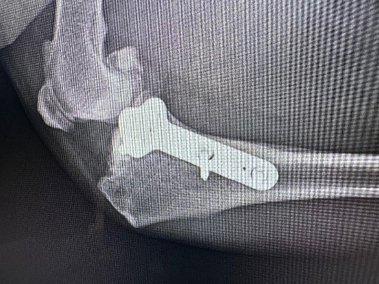
This is one of the preferred methods for early chronic cruciate disease to preserve remaining ligament fibers and prevent progression. It is also one of the preferred methods for large and giant breed dogs. This surgery involves cutting the tibia (shin bone), rotating it, and securing it with a bone plate so that the angle or slope of the top of the tibia that the femur (thigh bone) sits on is less steep. This prevents the tibia from sliding forward during walking/running. Bone healing generally takes 8-10 weeks. Care after surgery involves the pet wearing a plastic Elizabethan collar for 2 weeks to prevent licking of the incision, kennel rest with short leash walks to use the bathroom for 8-10 weeks, and possible bandaging of the limb. Potential complications are uncommon but include loosening of the implants, failure or delayed healing of the bone, and infection. Infection risk is increased if the pet is able to lick the suture line during the first 2 weeks after surgery, lack of compliance with bandage changes, or if the pet has bacteria elsewhere in their body (e.g. severe dental disease, UTIs, skin infections) that can then travel through the blood stream and settle on the implants. If infection is suspected, antibiotics, cultures, and eventual implant removal after bone healing has occurred may be necessary. TPLO surgery is not currently performed at TLC Animal Hospital. We currently recommend referral to a board certified surgeon for this procedure.
CBLO (CORA BASED LEVELING OSTEOTOMY)

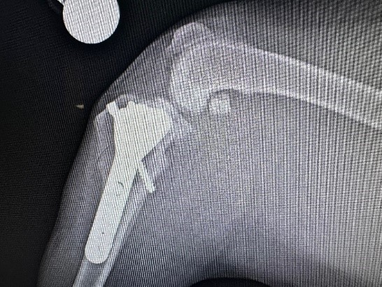
CBLO is a newer procedure than the TPLO, and has some advantages over the TPLO. CBLO is one of the preferred methods for early chronic cruciate disease to preserve remaining ligament fibers and prevent progression. It is also one of the preferred methods for large and giant breed dogs. Unlike the TPLO, this procedure can also be used in conjunction with a surgery to correct patellar alignment (for dogs with concurrent luxating patella), can be used in young dogs with open growth plates, and dogs with excessive tibial slope (when the top of the tibia is very steep). This surgery involves cutting the tibia (shin bone), rotating it, and securing it with a bone plate so that the angle or slope of the top of the tibia that the femur (thigh bone) sits on is less steep. This prevents the tibia from sliding forward during walking/running. The shape of the cut and bone plate are different from a TPLO. An additional compression screw helps to secure the two segments of bone, which allows for faster healing. Bone healing generally takes about 6 weeks. Care after surgery involves the pet wearing a plastic Elizabethan collar for 2 weeks to prevent licking of the incision, kennel rest with short leash walks to use the bathroom for 6 weeks, and possible bandaging of the limb. Potential complications are uncommon but include loosening of the implants, failure or delayed healing of the bone, and infection. Infection risk is increased if the pet is able to lick the suture line during the first 2 weeks after surgery, lack of compliance with bandage changes, or if the pet has bacteria elsewhere in their body (e.g. severe dental disease, UTIs, skin infections) that can then travel through the blood stream and settle on the implants. If infection is suspected, antibiotics, cultures, and eventual implant removal after bone healing has occurred may be necessary. CBLO surgery is offered at TLC Animal Hospital.
EXTRACAPSULAR REPAIR (LATERAL SUTURE)

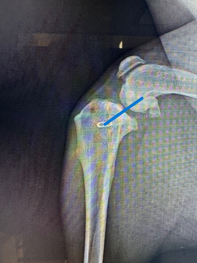
Extracapsular repair (also known as the lateral suture) involves placing a very strong suture around the knee to prevent forward motion of the tibia (represented by the blue line on the x-ray pictured). The suture will eventually fail, but if it can stay intact for at least 10 weeks, this gives the body enough time to lay down scar tissue to take over stabilizing the knee. This surgery does not spare any remaining cruciate ligament fibers or prevent progression of early cruciate disease. This technique works best for cats and small breed dogs. It can be used in large breed dogs, but has a higher rate of early implant failure in these pets. Extracapsular repair is offered at TLC Animal Hospital

- posted: Feb. 08, 2024
WHAT IS CRUCIATE DISEASE?

Canine knee joint – purple oval is the knee cap, blue line is orientation of the CrCL, pink wedge is the meniscus
In animals, the ACL is referred to as the Cranial Cruciate Ligament or CrCL. The CrCL is one of the ligaments important to stabilizing the knee and preventing the tibia (shin bone) from moving too far forward when walking/running. This ligament can either become suddenly torn due to injury (an acute tear, more common in small breed dogs and cats) or gradually fray and break down over time (chronic cruciate disease, more common in large breed dogs). Sometimes, the medial meniscus (one of the cushions in the knee) can also be torn, leading to more severe pain and lameness. For pets with acute CrCL tears, pain and lameness is usually sudden and severe. For dogs with chronic cruciate disease, they commonly only show “soreness” after strenuous exercise early in the disease. Over time, they either gradually become more and more sore and “slower” as joint inflammation and arthritis develops, or can become suddenly lame and painful if a large portion of the ligament or the meniscus suddenly tears. If left untreated, CrCL disease and injuries can cause significant arthritis, pain, and lameness. Once CrCL disease is affecting one leg, the pet will start weight shifting and putting more pressure on the other rear limb, increasing the likelihood of this leg also experiencing CrCL disease. It should be noted that in some animals with acute cruciate tears, their lameness may improve with time. This does not mean their ligament has healed or they don’t require surgery. Their comfort level initially improves as the joint swelling improves, but without intervention, arthritis will occur and cause worsening pain and lameness later on.
HOW IS CRUCIATE DISEASE DIAGNOSED?
Cruciate disease is often suspected and diagnosed with a combination of specific signs found on physical exam by a veterinarian and radiographs (x-rays) to evaluate arthritic changes and soft tissue inflammation in the knee joint. Definitive diagnosis is obtained by direct visualization of the CrCL ligament either by arthroscopy (inserting a small camera into the joint, generally performed by a surgical specialist) or by surgically exploring the joint (generally done during surgery to address the cruciate injury).
HOW IS CRUCIATE DISEASE TREATED?
Initially, your veterinarian should discuss efforts to protect the joints and reduce pain with appropriate pain medication, exercise restriction (no long walks or high impact activity), weight reduction (if needed to obtain a lean body condition), and introduction of good quality joint supplements (e.g. Dasuquin, Cosequin). Additional interventions, such as cold laser therapy, good quality omega fatty acid supplements (e.g. Antinol, Welactin), polysulfated glycosaminoglycan injections (e.g. Adequan), and physical therapy may be discussed. Joint supplements and weight control are recommended long term after a diagnosis of CrCL disease. If surgery is not pursued, pain medication and other interventions may also be required long term. Orthopedic braces are very rarely recommended.
The ideal treatment for cruciate disease is surgery to remove any meniscal tears, remove torn CrCL ligament fibers that contribute to joint inflammation, and stabilize the joint so that the tibia (shin bone) does not move forward during walking/running. Several surgical options are available. The most common are discussed here.
TPLO (TIBIAL PLATEAU LEVELING OSTEOTOMY)

This is one of the preferred methods for early chronic cruciate disease to preserve remaining ligament fibers and prevent progression. It is also one of the preferred methods for large and giant breed dogs. This surgery involves cutting the tibia (shin bone), rotating it, and securing it with a bone plate so that the angle or slope of the top of the tibia that the femur (thigh bone) sits on is less steep. This prevents the tibia from sliding forward during walking/running. Bone healing generally takes 8-10 weeks. Care after surgery involves the pet wearing a plastic Elizabethan collar for 2 weeks to prevent licking of the incision, kennel rest with short leash walks to use the bathroom for 8-10 weeks, and possible bandaging of the limb. Potential complications are uncommon but include loosening of the implants, failure or delayed healing of the bone, and infection. Infection risk is increased if the pet is able to lick the suture line during the first 2 weeks after surgery, lack of compliance with bandage changes, or if the pet has bacteria elsewhere in their body (e.g. severe dental disease, UTIs, skin infections) that can then travel through the blood stream and settle on the implants. If infection is suspected, antibiotics, cultures, and eventual implant removal after bone healing has occurred may be necessary. TPLO surgery is not currently performed at TLC Animal Hospital. We currently recommend referral to a board certified surgeon for this procedure.
CBLO (CORA BASED LEVELING OSTEOTOMY)

CBLO is a newer procedure than the TPLO, and has some advantages over the TPLO. CBLO is one of the preferred methods for early chronic cruciate disease to preserve remaining ligament fibers and prevent progression. It is also one of the preferred methods for large and giant breed dogs. Unlike the TPLO, this procedure can also be used in conjunction with a surgery to correct patellar alignment (for dogs with concurrent luxating patella), can be used in young dogs with open growth plates, and dogs with excessive tibial slope (when the top of the tibia is very steep). This surgery involves cutting the tibia (shin bone), rotating it, and securing it with a bone plate so that the angle or slope of the top of the tibia that the femur (thigh bone) sits on is less steep. This prevents the tibia from sliding forward during walking/running. The shape of the cut and bone plate are different from a TPLO. An additional compression screw helps to secure the two segments of bone, which allows for faster healing. Bone healing generally takes about 6 weeks. Care after surgery involves the pet wearing a plastic Elizabethan collar for 2 weeks to prevent licking of the incision, kennel rest with short leash walks to use the bathroom for 6 weeks, and possible bandaging of the limb. Potential complications are uncommon but include loosening of the implants, failure or delayed healing of the bone, and infection. Infection risk is increased if the pet is able to lick the suture line during the first 2 weeks after surgery, lack of compliance with bandage changes, or if the pet has bacteria elsewhere in their body (e.g. severe dental disease, UTIs, skin infections) that can then travel through the blood stream and settle on the implants. If infection is suspected, antibiotics, cultures, and eventual implant removal after bone healing has occurred may be necessary. CBLO surgery is offered at TLC Animal Hospital.
EXTRACAPSULAR REPAIR (LATERAL SUTURE)

Extracapsular repair (also known as the lateral suture) involves placing a very strong suture around the knee to prevent forward motion of the tibia (represented by the blue line on the x-ray pictured). The suture will eventually fail, but if it can stay intact for at least 10 weeks, this gives the body enough time to lay down scar tissue to take over stabilizing the knee. This surgery does not spare any remaining cruciate ligament fibers or prevent progression of early cruciate disease. This technique works best for cats and small breed dogs. It can be used in large breed dogs, but has a higher rate of early implant failure in these pets. Extracapsular repair is offered at TLC Animal Hospital
Office Hours
Monday
8:00 AM - 5:30 PM
Tuesday
8:00 AM - 5:30 PM
Wednesday
8:00 AM - 5:30 PM
Thursday
8:00 AM - 5:30 PM
Friday
8:00 AM - 5:30 PM
Saturday
8:00 AM - 12:00 PM
Sunday
Closed
Walk In Hours
in case of emergency or an urgent health need after posted walk in times, please call or consult with the front desk to check doctor availability
Monday
8:00 am - 4:00 pm
Tuesday
8:00 am - 4:00 pm
Wednesday
8:00 am - 4:00 pm
Thursday
8:00 am - 4:00 pm
Friday
8:00 am - 4:00 pm
Saturday
8:00 am - 10:00 am
Sunday
Closed


